

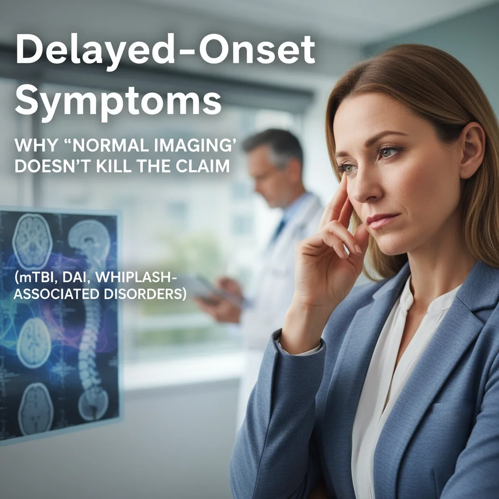
Delayed-Onset Symptoms: Why "normal imaging" doesn't kill the claim (mTBI, DAI, whiplash-associated disorders)
The absence of abnormal findings on conventional neuroimaging represents one of the most significant challenges in establishing causation for traumatic brain injury claims, particularly those involving mild traumatic brain injury (mTBI), diffuse axonal injury (DAI), and whiplash-associated disorders. Defense counsel and insurance adjusters frequently exploit normal CT scans and MRI findings to contest the validity of claims, operating under the misconception that structural imaging abnormalities constitute prerequisite evidence for brain injury. This perspective fundamentally misunderstands both the pathophysiology of these conditions and the inherent limitations of current imaging modalities in detecting subtle but functionally devastating neural injuries.
The Pathophysiology of Delayed-Onset Neurological Manifestations
The temporal evolution of traumatic brain injury symptoms follows a complex cascade of neuroinflammatory, metabolic, and structural changes that may not become clinically apparent for weeks, months, or even years following the initial trauma. Primary injury mechanisms, including acceleration-deceleration forces and rotational shearing, initiate secondary injury cascades involving excitotoxicity, calcium dysregulation, mitochondrial dysfunction, and neuroinflammation. These processes continue to evolve long after the initial traumatic event, producing progressive axonal degeneration, synaptic dysfunction, and network disruption that conventional imaging techniques cannot adequately capture.
Diffuse axonal injury, in particular, represents a microscopic pathological process characterized by widespread disruption of white matter tracts throughout the brain. The mechanical forces that produce DAI cause immediate stretching and delayed secondary axotomy of nerve fibers, resulting in progressive Wallerian degeneration that may continue for months following the initial injury. This delayed pathological progression explains why cognitive, behavioral, and physical symptoms may not manifest until weeks or months after trauma, creating apparent temporal gaps that defense teams exploit to challenge causation.
Limitations of Conventional Neuroimaging in Detecting Subtle Brain Injury
Standard neuroimaging protocols, including non-contrast CT scanning and conventional MRI sequences, possess significant limitations in detecting the microstructural abnormalities characteristic of mTBI and DAI. CT scanning, while excellent for identifying hemorrhage, mass effect, and fractures, lacks the resolution necessary to visualize microscopic axonal injury, subtle edema, or early ischemic changes. The sensitivity of CT for detecting mild traumatic brain injury ranges from 4% to 8%, rendering normal CT findings essentially meaningless in the context of mTBI evaluation.
Conventional MRI sequences, though superior to CT in detecting subtle abnormalities, remain inadequate for comprehensive assessment of diffuse axonal injury. Standard T1-weighted, T2-weighted, and FLAIR sequences may demonstrate normal findings even in the presence of significant functional impairment. The absence of signal abnormalities on conventional MRI does not exclude the presence of axonal injury, particularly in cases where the pathological changes occur at the cellular level without producing sufficient tissue water content alterations to generate signal changes on routine sequences.
Advanced Neuroimaging Modalities and Their Clinical Utility
Diffusion tensor imaging (DTI) represents the most promising advancement in detecting microstructural brain injury, providing a quantitative assessment of white matter integrity through measurement of water diffusion properties. DTI parameters, including fractional anisotropy (FA) and mean diffusivity (MD), can identify subtle alterations in white matter microstructure that correlate with functional deficits in patients with normal conventional imaging. Studies consistently demonstrate that DTI abnormalities may persist for months to years following mTBI, providing objective evidence of ongoing pathological processes in patients with normal structural imaging.
Functional MRI (fMRI) and magnetic resonance spectroscopy (MRS) provide additional insights into the neurochemical and functional consequences of brain injury. fMRI can identify alterations in neural network connectivity and activation patterns that correlate with cognitive and behavioral symptoms, while MRS detects metabolic abnormalities, including reduced N-acetylaspartate levels, elevated choline, and increased lactate concentrations. These advanced modalities frequently reveal abnormalities in patients with normal conventional imaging, providing crucial objective evidence supporting symptom validity.
Clinical Presentation Patterns in Delayed-Onset Syndromes
The clinical manifestations of delayed-onset neurological symptoms follow predictable patterns that reflect the underlying pathophysiology and anatomical distribution of injury. Cognitive symptoms, including attention deficits, memory impairment, and executive dysfunction, typically emerge as the most prominent delayed features, often becoming apparent only when patients return to cognitively demanding activities. These deficits may not manifest during the acute hospitalization period when patients remain in structured, low-demand environments.
Behavioral and psychiatric symptoms frequently demonstrate the most delayed onset, with depression, anxiety, irritability, and personality changes emerging weeks to months following injury. The delayed appearance of these symptoms reflects the complex interaction between structural brain injury, psychological adaptation, and environmental stressors, creating multifactorial causation scenarios that require sophisticated medical-legal analysis.
Whiplash-Associated Disorders: Beyond Cervical Spine Imaging
Whiplash-associated disorders encompass a spectrum of symptoms extending beyond simple cervical spine injury to include complex interactions between cervical spine dysfunction, vestibular disturbance, and central nervous system sensitization. Normal cervical spine imaging, including X-rays, CT scans, and MRI studies, does not exclude the presence of significant functional impairment resulting from ligamentous injury, facet joint dysfunction, or cervical disc pathology below the resolution threshold of conventional imaging.
The pathophysiology of whiplash-associated disorders involves complex mechanisms, including cervical spine ligamentous injury, facet joint capsule disruption, and potential brainstem or upper cervical cord injury resulting from excessive cervical hyperextension and hyperflexion. These injuries may not produce structural abnormalities visible on routine imaging while causing significant functional impairment through nociceptive sensitization, vestibular dysfunction, and autonomic dysregulation.
Documentation Strategies for Delayed-Onset Claims
Establishing causation for delayed-onset symptoms requires comprehensive documentation strategies that capture the temporal evolution of symptoms and their functional impact. Medical records must demonstrate a clear timeline linking the traumatic event to the gradual emergence of symptoms, including detailed documentation of the patient's pre-injury functional status and the progressive deterioration following trauma.
Neuropsychological testing provides crucial objective evidence of cognitive dysfunction that may not be apparent on clinical examination. Serial neuropsychological evaluations can document the evolution of cognitive deficits over time, providing objective evidence of progressive impairment that correlates with the expected pathophysiology of traumatic brain injury. These assessments must be conducted by qualified neuropsychologists using validated testing batteries appropriate for the patient's educational and cultural background.
Functional Assessment and Activities of Daily Living Documentation
The documentation of functional impairment through activities of daily living (ADL) assessments provides essential evidence supporting delayed-onset symptom claims. These assessments must capture not only the patient's subjective reports but also objective observations from family members, employers, and healthcare providers who can document changes in the patient's functional capacity.
Occupational therapy evaluations, vocational assessments, and disability rating scales provide standardized measures of functional impairment that can be tracked over time to demonstrate progressive deterioration or improvement. These assessments must be conducted at regular intervals to capture the evolution of functional capacity and establish the temporal relationship between injury and functional decline.
Medicolegal Implications and Expert Testimony Requirements
The successful prosecution of delayed-onset symptom claims requires expert testimony that can educate juries about the complex pathophysiology underlying these conditions and the limitations of conventional imaging in detecting subtle brain injury. Expert witnesses must be prepared to explain why normal imaging findings do not exclude the presence of significant brain injury and how delayed-onset symptoms represent expected consequences of the underlying pathophysiology rather than evidence against causation.
Medical experts must demonstrate familiarity with the current scientific literature regarding delayed-onset symptoms and advanced neuroimaging techniques. They must be prepared to distinguish between expected delayed manifestations of traumatic brain injury and unrelated medical conditions that may coincidentally develop following trauma. This requires comprehensive review of the patient's medical history, careful analysis of symptom patterns, and consideration of alternative explanations for the observed clinical findings.
The integration of advanced neuroimaging findings with clinical presentation and functional assessment data provides the strongest foundation for establishing causation in delayed-onset symptom cases. Medical-legal consultants must work closely with treating physicians to ensure appropriate advanced imaging studies are obtained and interpreted by qualified specialists familiar with traumatic brain injury pathophysiology.
Understanding the complex relationship between traumatic brain injury pathophysiology and the limitations of conventional imaging represents essential knowledge for legal professionals handling these challenging cases. Normal imaging findings should never be accepted as definitive evidence against brain injury, particularly in cases involving delayed-onset symptoms consistent with the expected evolution of mTBI, DAI, or whiplash-associated disorders.